- HOME
- ABOUT US
- PRACTITIONERS
- PATIENTS
- CONDITIONS
- Skin & Nail Fungal Infections Protocol
- Chronic Kidney Disease (CKD) Protocol
- Fatty Liver Disease & Hepatitis Protocol
- Thyroid Disorders Protocol
- Vitiligo Protocol
- Sprains and Strains
- C. Difficile
- Dupuytren Contracture
- Urinary Incontinence & Pelvic Organ Prolapse Protocol
- Arrhythmia and Afib Protocol
- Telomere +
- WeiDetox
- ATP +
- Sarcoidosis Protocol
- Sinusitis and Nasal Congestion Protocol
- Anemia
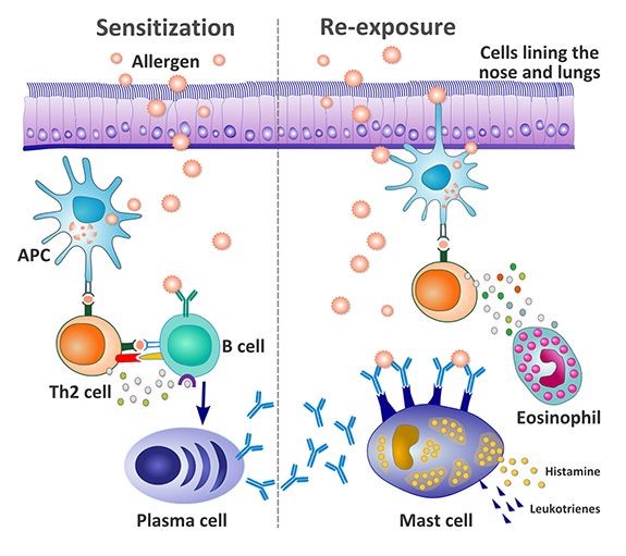
- Food Allergies and Intolerances
- Diverticulitis
- Eczema & Atopic Dermatitis
- Male Infertility
- Erectile Dysfunction (ED) Protocol
- Asthma Protocol
- Cataracts Protocol
- Hair Loss Protocol
- Stress, Anxiety, PTSD and Depression
- Long COVID Symptoms
- Menopause
- Atherosclerosis
- Stroke and Post-Stroke Recovery
- Hyperlipidemia (High Cholesterol)
- Female Infertility
- Heart Inflammation
- Heavy Metal Toxicity
- Stenosis and Bone Spur
- PMS, Post Menstrual Syndrome, & Premenstrual Dysphoric Disorder
- Uterine Fibroids & Ovarian Cysts
- Teeth & Gums Protocol
- Weight Control
- Lyme Disease
- Macular Degeneration
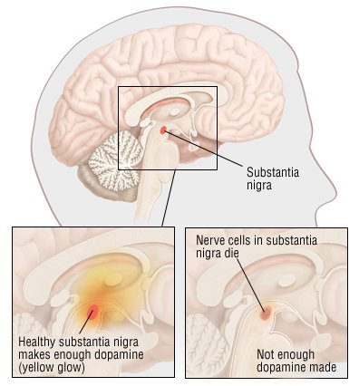
- Parkinson's Disease
- Hip Pain Protocol
- Acid Reflux and GERD
- Gallbladder Stone
- Nervous System Viral Infections and Complications
- Cyst Protocol
- Cold and Flu
- Low Testosterone
- Acne Protocol
- Osteoarthritis and Bone-on-Bone Protocol
- Sleep Apnea Protocol
- Ankylosing Spondylitis Protocol
- Bulging and Herniated Disc Protocol
- Glaucoma & Cataracts Protocol
- Allergies, Asthma, and Eczema
- Migraine and Headache
- Ear Infection & Tinnitus
- Insomnia
- Attention Deficit Hyperactivity Disorder (ADHD)
- Concussion
- Treatment Sequence
- Urinary Tract Infection Protocol
- Cystic Fibrosis
- Kidney Stone
- Stress, Anxiety, and Depression
- High Blood Pressure
- Hernia
- Gout
- Cardiovascular Disease
- Men's Health
- Bursitis
- Prostate Enlargement
- Lupus
- Thyroid Disorders
- TMJ Syndrome
- Hashimoto's Thyroiditis
- Bone Spur
- Baker's Cyst
- Bronchiectasis
- IBS
- Fibromyalgia
- Liver Cirrhosis
- Frozen Shoulder & Rotator Cuff Injury
- Cancer Care
- Pulmonary Fibrosis & Interstitial Lung Disease
- Avascular Necrosis & Piriformis Syndrome
- Viral Infections
- Women's Health
- Type II Diabetes
- Psoriasis and Psoriatic Arthritis
- Osteoarthritis Customized Treatment
- Leaky Gut
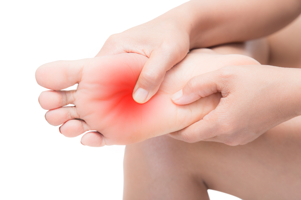
- Peripheral and Foot Neuropathy
- IBD, Ulcerative Colitis, Crohn's Disease
- Tendinosis, Tendinitis, Tendon Tear and CTS
- Sinusitis
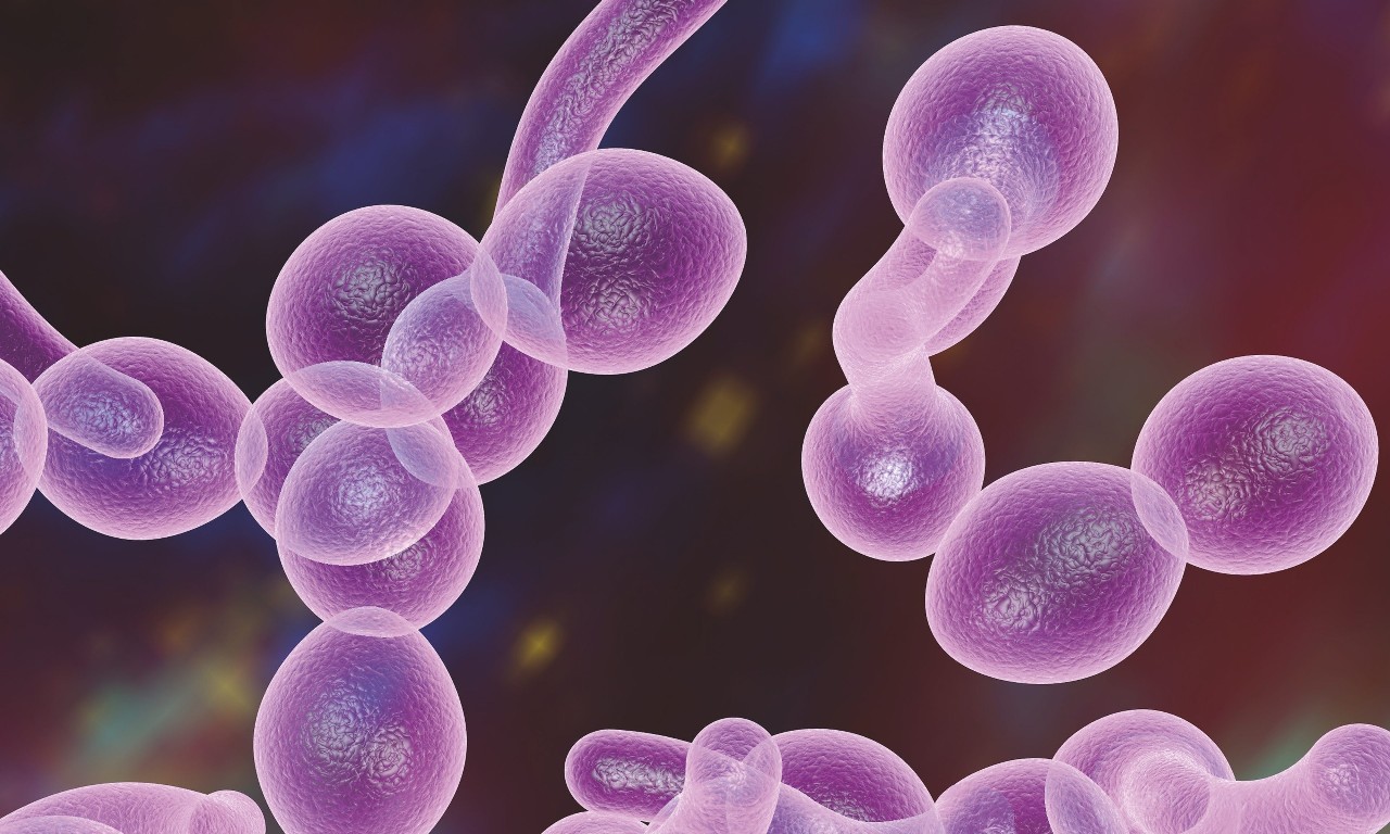
- Candida & Fungal Infections Protocol
- COPD, Emphysema, and Chronic Bronchitis Protocol
- Rheumatoid Arthritis
- Meniscus and Cartilage Tear
- PRODUCTS
- CONTACT US